"It never gets easier, you just go faster." - Greg Lemond
Showing posts with label disease. Show all posts

Friday, July 12, 2013

Gluten intolerance and neurological illness

Introduction
Gluten containing grains (wheat, rye, barley) make up a large portion of our daily diet, and these grains have become staple foods in the Western world.  Gluten itself is a protein structure that has been shown to elicit an immune response in some susceptible individuals.  Those who are gluten intolerant can be afflicted with what is known as celiac disease in which an immune response against self-enzymes is elicited upon ingestion of even tiny amounts of gluten (1).

Adapted from (6)

Celiac disease is a systemic immunological disease that is most commonly associated with gut dysfunction and gastointestinal symptoms.  Celiac disease is becoming an increasingly commonly recognized disorder, and it is estimated that approximately .75-1.0% of the general population suffers from some degree of celiac disease (1,2).  While the traditional concept is that celiac disease results in gastrointestinal distress, there appear to be a number of symptoms that occur outside of the gastointesinal system.  However, an interesting symptom commonly found in persons who suffer from gluten intolerance or celiac disease are unexplained neurological symptoms. This is actually not that new of a concept, as early as 1966 doctors have associated celiac disease with neurological illness (3).

Background
The biological foundation for this observation is perhaps best evidenced by the fact that the gastrointestinal system actually has its own nervous system which communicates directly with the central nervous system.  There has since been the hypothesis proposed that states:  "Gluten causes symptoms in both celiac disease and non-celiac gluten intolerance, by its action on the nervous system" (1).

Proper neurological function is required for general health and well being; there has been a good amount of investigation into the relationship between gluten sensitivity and neurological function.  Proper digestive activity, including gut motility and gall-bladder function rely on proper nervous system activity.  Thus, the gastrointestinal symptoms reported in celiacs could largely be related to nervous system dysfunction (1,3).   In a report investigating the occurrence of neurological disease in celiacs and non-celiacs it was found that 51.4% of those with celiac suffered some sort of neurological dysfunction, while only 19.9% of those without celiac presented with neurological illness (1).  Other studies paint a slightly less dire picture, with between 10-25% of patients with confirmed celiac presenting with neurological illness of some sort, however the neurological dysfunction that is associated with celiac disease still presents a  large economic and social burden (2).  Similarly, there is a high prevalence (46%) of headache/migraines found in patients with celiac disease compared to those without celiac (29%) (1,4,5).  Taken together, these data suggest that those with celiac are over two-times more likely to have neurological dysfunction, of some sort.

In studies conducted at the Mayo clinic, patients with childhood onset celiac disease suffered from ataxia, peripheral neuropathy and seizures at an abnormally high rate (1).  Dementia has been associated with adult-onset celiac disease, such that dementia began soon after the onset of celiac symptoms, although this appears to be a rather rare phenomenon.  Furthermore, in patients in which celiac disease is associated with neurological dysfunction, cognitive impairment is commonly reported as well as amnesia (1).  The onset of cognitive impairment is also especially found to begin soon after the onset of celiac disease-like symptoms (1).

Adapted from (6)

One of the more interesting concepts regarding gluten sensitivity and gluten tolerance is the differentiation between celiac disease and gluten sensitivity.  I have more or less lumped these two terms together there, which I believe is a practical consideration as our diagnostic methods for these different conditions are constantly evolving and have yet to been unified.  For example, Hoffenberg et al. reported on a large cohort of children who were screened for evidence of celiac disease and found that 724 presented with high anti-gliadin levels (a diagnostic measure of celiac) (4).  However, upon histological examination of these children, 4.3% were define celiac (94% improvement with gluten free diet), 6.6 % were possible celiacs (with 75% improvement on a gluten free diet) and 89.1% were not-celiacs, despite the fact that 53% of these children reported improvement of symptoms with a gluten free diet (1,4).

Interestingly and importantly, neurological symptoms (tiredness, lethargy, irritability, sleep disturbance) were commonly reported in all three of these groups with 71% of the confirmed celiacs, 65% of the possible celiacs, and 51% of the non-celiacs reporting these symptoms (1).

While the association between celiac disease and neurological dysfunction appears to be fairly strong, without understanding of potential mechanisms, I remain unconvinced.  However, there does appear to be a fairly strong mechanistic basis for these observations.  Celiac is an immunological disorder, in which B cell produce auto-antibodies against transglutaminase or gliadin, the enzymes required to digest and absorp gluten or a portion of wheat, respectively (6).  Thus, the current belief is that gluten sensitivity results in auto-antibody production, which in turn damages nerve cells and results in inflammation.  Evidence for this hypothesis consists of the fact that 64% of celiacs who presented with neurological disease also had anti-ganglioside (a nerve cell) antibodies (5,6), in their circulation.  These antibodies have been shown to bind to a number of critical nerve sites that result in immune mediated damage to the nerve.  

Furthermore, studies mapping blood flow in the brains of patients who are gluten intolerant who are on a gluten free diet and those who are not have found that there is significant differences in the perfusion of various areas of the brain between the two groups (7).  While the direct cause of this is unclear, this could be related to inflammation and immunoreactivity, or direct toxicity from gluten.

Conclusions
While alterations in growth and body composition may be expected in persons suffering from gastointestinal illness such as celiac, the concept that this is accompanied by neurological illness as well has significant implications and is possibly less appreciated.  Those with celiac may also suffer from a wide spectrum of neurologic an psychiatric symptoms, including neuropathy, ataxia, migraines, epilepsy, lethargy, irritability, and depression (4,5). 

A search of the literature suggests that gluten can cause neurological harm through a combination of cross reacting antibodies, as well as further immunological damage, or toxicity (6),7. 

Works Cited

1)  Ford RP. The gluten syndrome: a neurological disease. Med Hypotheses 2009;73(3):438–40.

2)  Currie S, Hadjivassiliou M, Clar MJ, et al.  Should we be ‘nervous’ about coeliac disease? Brain abnormalities in patients with coeliac disease referred for neurological opinion.  J Neurol Neurosurg Psychiatry 2012;83:1216–1221 

3)  Cooke W, Smith W. Neurological disorders associated with adult coeliac disease. Brain 1966;89:683–722.

4) Hoffenberg EJ, Emery LM, Barriga KJ, et al.  Clinical features of children with screening identified evidence of celiac disease.  Pedatrics 2004; May;113(5):1254-9. 

5)  Bushara KO.  Neurologic Presentation of Celiac Disease.  Gastroenterology 2005;128:S92–S97
  
6) Green PHR, Alaedin A, Sander HW, et al.  Mechanisms underlying celiac disease and its neurological manifestations.  Cell. Mol. Life Sci. 2005; 62

7)  Addolorato G, Di Giuda D, De Rossi G, et al. Regional cerebral hypoperfusion in patients with celiac disease. Am J Med 2004;116:312–7.

Tuesday, March 26, 2013

Salt Matters


Introduction
 The health outcomes associated with a high salt diet are generally negative, and include increased risk of cardiovascular disease risk, stroke, and kidney disease (1).  Because salt intake influences the total osmolarity of your blood, increased consumption of salt, aka sodium-chloride (NaCl), rich foods results in the body shunting more fluid to the blood, which requires various compensatory mechanisms, including increased workload on the cardiovascular system (2).  There is substantial evidence that those at risk for cardiovascular disease due to high blood pressure should reduce NaCl intake.  However, recent high impact studies have been published that demonstrate salt intake may also influence the progression of other diseases.
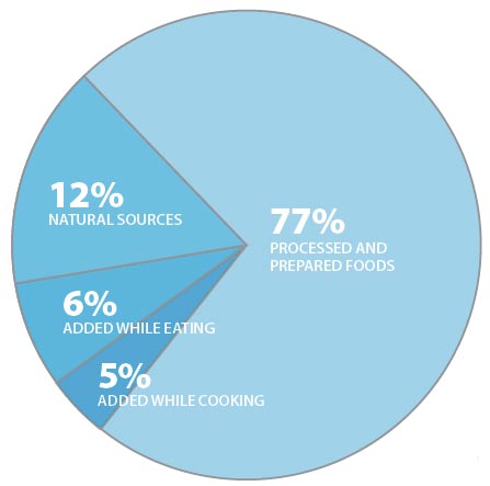
Salt sources in American diet (source NIH)
There has been a significant increase in the incidence and frequency of autoimmune disease in the American population over the last half-century (3).  Examples of these diseases include inflammatory bowel disease, rheumatoid arthritis, type I diabetes, multiple sclerosis, and lupus erythematosis.  In order to understand whether certain environmental factors may be responsible for the increased incidence of autoimmune disorders in the United States, researches have examined the relationship between changes in the Western diet and immune function.  Increased consumption of processed foods or “fast” foods containing copious amounts of NaCl has represented a significant dietary change over the past half-century (4), although the relationship between high NaCl intake and autoimmune disease has not been well characterized.

Cutting Edge
Recent studies published in Nature Letters have provided a solid foundation for future research focused on the relationship between NaCl intake and autoimmune immune disease.  A recently discovered immune cell, known as a Th17 T cell, has been suggested to play a role in the etiology of numerous autoimmune and proinflammatory diseases.  Th17 cells are known to heavily participate in chronic inflammatory diseases as well as hyperactivation of the immune system during immune responses to various pathogens such as influenza (5).  Because these cells are mediators of inflammation, they play a critical role in protecting the host from infectious agents, however, excessive activation of the immune system resulting in chronic inflammatory or hyperinflammatory responses has potentially lethal consequences to the host.

Thus under normal circumstances, Th17 cells are tightly regulated immune cells that are present at relatively low frequencies in the body.  In has been demonstrated that Th17 cells can be found at increased numbers during autoimmune disease, and deletion of Th17 cells using genetic ablation reduces the severity of autoimmunity (5, 6).  These observations reinforce the notion that Th17 cells are critical mediators of autoimmune disease.  Because our understanding of these cells is in its infancy, it is not quite understood what factors determine the production of Th17 cells.  However, among various cytokines, the inflammatory cytokine, interleukin 23 (IL-23), has been shown to stabilize and reinforce Th17 development (7).  

In two very timely publications, scientists show that high sodium (Na) plays a role in the development and pathogenic nature of Th17 cells.  Studying the molecular aspects of Th17 cell differentiation, Wu et al. show high Na concentrations induce the activation of signaling within T cells that supports development into Th17 cells (8).  This was shown to be related to the activation of an intracellular protein known as serum glucocorticoid kinase 1 (SGK1), a salt-sensitive protein, meaning its activation is highly dependent on salt concentrations.  Activation of SGK1 in T cells results in increased sensitivity to IL-23, which as mentioned earlier supports Th17 development.  

In the vary same issue of Nature Letters Kleinewietfeld et al. discovered culture of human T cells in media containing increasing NaCl concentrations promoted the development of Th17 cells in a dose dependent fashion (9).  Using an “experimental” model of autoimmunity, the authors of this study fed mice that were destined to develop autoimmune encephalitis (an autoimmune disease similar to multiple sclerosis) a high salt diet and found that mice fed a high NaCl diet presented with more severe autoimmunity than mice fed a normal NaCl diet (9).  Further studies by Wu et al. found that mice fed a high salt diet alone had marked increase in the frequency of Th17 cells in gut associated lymphoid tissues, and that mice harboring a genetic deletion of SGK1 had reduced Th17 cell development in response to a high salt diet.  Perhaps even more important, Wu et al. present data in agreement with Kleinewietfeld et al. in that mice fed a high salt diet exhibited more severe autoimmunity than mice fed a normal salt diet and that this severe autoimmunity was SGK1 dependent.  

Conclusions
Ok, great, but what does this all mean? 

I’m sure the media will blow most of this out of proportion... (Foxnews).   Do these studies say that having a high salt intake will result in autoimmune disease?  NO. 

These are experimental models of autoimmunity, in which mice are immunized with proteins that forcefully induce autoimmunity.  However, these two studies form an attractive hypothesis supporting the role of the Western diet in immune dysfunction.  Furthermore, these data provide framework for future studies examining the direct role of NaCl intake on the incidence of autoimmunity, especially in individuals who might be genetically predisposed to developing autoimmune disease.

What can be taken away from these studies is that there is some evidence that excess salt in the diet can alter immune cell development, which has dangerous implications if one is predisposed to developing autoimmunity (such as in these mice).  The authors conclude that determining whether a “true low-salt diet, representing the conditions in which Homo sapiens were environmentally selected in Africa” has the potential to reduce risk of autoimmunity will be very difficult because of Western culture, which to me is a truly disappointing conclusion (obviously they’ve never heard of Paleo!).  Instead the authors suggest looking for pharmacological agents that block the molecular pathways described above (yay, just give people more drugs and lets continue to eat like crap!).  I think based on these studies there is real evidence that high salt diets have the potential to further exacerbate inflammatory responses which are clearly detrimental to human health.   

Works Cited

1.         Brown IJ, Tzoulaki I, Candeias V, Elliott P. Salt intakes around the world: implications for public health. Int J Epidemiol. 2009 Jun;38:791-813.
2.         Appel LJ, Frohlich ED, Hall JE, Pearson TA, Sacco RL, Seals DR, Sacks FM, Smith SC, Jr., Vafiadis DK, Van Horn LV. The importance of population-wide sodium reduction as a means to prevent cardiovascular disease and stroke: a call to action from the American Heart Association. Circulation.  Mar 15;123:1138-43.
3.        Ascherio A, Munger KL. Environmental risk factors for multiple sclerosis. Part II: Noninfectious factors. Ann Neurol. 2007 Jun;61:504-13.
4.         McGuire S. Institute of Medicine. 2010. Strategies to Reduce Sodium Intake in the United States. Washington, DC: The National Academies Press. Adv Nutr.  Nov;1:49-50.
5.         Korn T, Bettelli E, Oukka M, Kuchroo VK. IL-17 and Th17 Cells. Annu Rev Immunol. 2009;27:485-517.
6.         Lee Y, Awasthi A, Yosef N, Quintana FJ, Xiao S, Peters A, Wu C, Kleinewietfeld M, Kunder S, et al. Induction and molecular signature of pathogenic TH17 cells. Nat Immunol.  Oct;13:991-9.
7.         Aggarwal S, Ghilardi N, Xie MH, de Sauvage FJ, Gurney AL. Interleukin-23 promotes a distinct CD4 T cell activation state characterized by the production of interleukin-17. J Biol Chem. 2003 Jan 17;278:1910-4.
8.         Wu C, Yosef N, Thalhamer T, Zhu C, Xiao S, Kishi Y, Regev A, Kuchroo VK. Induction of pathogenic T17 cells by inducible salt-sensing kinase SGK1. Nature.  Mar 6.
9.         Kleinewietfeld M, Manzel A, Titze J, Kvakan H, Yosef N, Linker RA, Muller DN, Hafler DA. Sodium chloride drives autoimmune disease by the induction of pathogenic T17 cells. Nature.  Mar 6.